Disease Management
BLUE TONGUE (BT)
This is acute infectious but not contagious disease of sheep
characterized by fever, inflammation and ulceration of buccal mucosa and
tongue.
It is basically a disease of sheep and young sheep within the age group
of one year are more prone to infection. Suckling lambs are relatively
resistant due to their acquired passive immunity through colostrum.
The disease occurs mainly during the rainy season particularly in the
months of October, November and December.
Causes
-
It is caused by Arthropod-borne orbi virus in the family of
Reoviridae.
-
Mosquitoes and other ectoparasites like sheep ked, Melophagus ovinus
may transmit the disease mechanically.
-
The disease is more prevalent in late summer and early autumn which
makes conducive environment for the multiplication of the vectors.
- Transmission through semen and placental route is possible.
-
The virus is resistant to decomposition, desiccation and against
antiseptic agents.
Clinical symptoms
- Fever, depressed attitude and off feed.
-
Reddening and swelling of nose and oral mucosa, profuse nasal and oral
discharge
-
Inflammation and ulceration of lips, gums, buccal mucosa and tongue,
Cyanotic (bluish) appearance of tongue, tilting of neck towards one
side
- Lameness, reddening and swelling of coronary band of the limbs.
-
Congestion of conjunctival mucous membranes and matting of eyelids,
- Foul smelling diarrhoea.
- Dyspnoea, snoring and Pneumonia may be observed.
- Death due to respiratory failure.
General prevention and control measures
-
Separation of sick animals should be made and affected animals should
be kept away from solar exposure.
-
Adequate rest to the affected animal and should be fed with porridge
made of rice and ragi.
-
Apply glycerin or animal fat on the ulcers as first aid and contact
the veterinarian immediately.
-
Ulcers in the mouth can be treated with saline water or dissolve 1g of
Potassium permanganate in 1 liter of water and wash the mouth 2 to 3
times a day with this solution.
- Animals should not be allowed for grazing.
- Proper Vaccination of animals with regular intervals.
-
Vaccination schedule: First vaccination at 3 months of age and next
vaccination once in a year.
-
Attempt should be made to control the vector (culicoides) population
with fly repellants.
-
Use of ectoparasiticides injections should be suggested in the areas
more prone to vector population.
-
Grazing of the animals should be avoided in areas where there is lot
of vectors.
-
Cattle may act as carrier. Viraemic stage remains in them for more
than 5 weeks. So movements of cattle should be restricted.
-
Importation of animals from the areas prevailing the disease should be
avoided.
-
Strict regulation is to be followed to prevent entry of diseased
animals from endemic zones.
-
The spread of the disease can be controlled by the use of insect
repellents, external application of fly repellents and spraying of
butox (1ml in 1 liter of water) in the breeding places of the insects.
- The sheep can be housed in insect proof sheds.
-
Cloud of smoke with dried leaves / wood during 6 - 8 P.M. might help
to keep off Culicoides from sheep sheds.
SHEEP-POX
About this disease
-
It is an acute to chronic viral disease of sheep and goats
characterized by generalized pox lesions throughout the skin and
mucous membranes.
-
All breeds of sheep and goats irrespective of age and sex are
affected.
-
It is possible to infect goats with sheep pox virus and sheep with
goat pox virus.
-
Sheep are naturally susceptible to sheep pox. Younger sheep are more
susceptible over old ones.
- Disease occurrence period is April- June.
-
Sheep-pox is highly contagious disease which may cause a mortality of
20 to 50 per cent in animals below the age of 6 months, and causes
damage to the wool and skin in adults.
-
Of the pox diseases, Sheep-pox ranks only second to human small-pox in
virulence.
-
The disease is transmissible to in-contact goats but not to other
species of animals. It, however, spreads slowly.
Causes
-
It is caused by a member of the genus Capri pox virus, pox viridae
family.
-
Cutaneous lesions (crust, nodules) resulting in aerosols, saliva,
faeces, nasal secretions from sick animals for 1-2 months and dried
scabs at ambient temperature may be the source for spread of virus.
-
Susceptible to highly alkaline or acid PH and virus remains viable for
as long as six months.
- Virus is susceptible to 560c for 2 hrs and 650c for 30 minutes.
-
The usual mode of transmission is from direct contact with the
infected animal.
-
Indirect transmission by contaminated litter, fodder, water and
attendants may spread the virus through mechanical ways.
- The virus may gain entrance through wound and abrasions.
-
The virus may be present in skin papules. While the affected animals
rub their body on other animals, the virus is passed directly to
susceptible animals.
-
The biting insects (mechanical vectors) may inoculate the virus
intradermaly or subcutaneously.
- Aerosol or droplet infection is quite possible.
-
Dog, cat etc. may mechanically transport the virus to other places.
-
The virus may pass from infected mother to the foetus through
placenta.
Symptoms
-
The disease is characterized by high fever, and symptoms of pneumonia
and acute enteritis.
-
Skin lesions appear particularly in parts free from wool, notably
around the eyes, inner side of the thigh, udder and under surface of
the tail.
-
Skin papules appear in 2-5 days following temperature and first appear
on the hairless parts of the skin.
-
Papules like pock lesions appear in all the parts of the body, e.g.,
lips, cheeks, snout, nostril, face, ear, feet, thigh, abdomen, eye
lid, neck, teat and udder.
-
The internal organs such as trachea, lungs, kidneys and intestines are
also affected.
-
The disease results in emaciation and, as already mentioned, frequent
deaths of affected animals.
-
The eyelids are swollen and they may completely cover the eye ball.
- Mucopurulent discharges from eyes and nose.
-
Animals become weak, disoriented and eventually unable to stand.
-
The mucous membrane of the eyes, nose, lips, vulva and prepuce become
necrotic.
-
Animals die due to the development of labored breathing as a result of
broncho-pneumonia.
-
Animals that survive develop scab and shed over a period of 3-6 weeks,
leaving a raw granulating area.
Treatment, Prevention and Control
-
The diseased animal should be treated with palliatives. In the young
ones, nursing is more important than medication.
-
The infected litter should be burnt and the bedding changed every day.
Affected animals should be kept on soft diet.
-
The ulcers on the skin should be washed with potassium permanganate
lotion and dusted with boric acid; strict hygienic measures should be
adopted.
-
The method of control by the use of vesicular fluid was in vogue for
dealing with sheep-pox. A couple of sheep may be first inoculated with
the vesicular fluid on the under surface of the tail or the inner side
of the ear by scarification.
-
In about 4 to 6 days, vesicles appear at the spot, and the fluid
collected from these vesicles, mix with equal parts of glycerol,
served as a vaccine.
-
Vaccination may be done by scarification inside the ear or under the
tail. In about 15 to 20 days, the animals become resistant to the
disease.
-
Isolation of infected herds and sick animals for at least 45 days
after recovery.
-
Use of disinfectants like ether (20%), chloroform and formalin (1%),
phenol (2%) to prevent the transmission of disease.
-
Isolation of infected herds and sick animals for at least 45 days
after recovery.
- Quarantine before introduction into herds.
- Proper disposal of cadavers and products.
-
First vaccination at 3months of age and Next vaccination is once in
year. Normally vaccinated in the month of Feb-March
BRUCELLOSIS
Transmission
- A large number of organisms are eliminated ruing abortion.
- The mode of entry is by ingestion or via conjunctiva.
-
The aborted foetus, vaginal discharge and milk from infected goats
contain a large number of organisms.
Symptoms
-
In infected goats and sheep, state of abortion may occur followed by a
quiescent period during which a few abortions occur.
- The aborted animals do not breed.
-
After 2 years or more another abortion storm is likely to occur.
Diagnosis, Treatment and Control
-
It is not possible to diagnose brucellosis on the basis of symptoms
alone.
-
The suspicion is aroused when humans in contact suffer from undulant
fever and there is poor breeding record in shepherd and evidence of
mastitis.
-
The diagnosis can be done by isolation of organisms and by serological
tests.
TETANUS
-
This is an infectious, non-febrile disease of animals and man, and is
characterized by spasmodic tenancy and hyperesthesia.
-
It is caused by bacterial toxin characterized by spasmodic contraction
of skeletal muscles.
- Sheep and goat are more susceptible than cattle.
- This disease is prevalent all over the world.
Causes
-
The disease is caused by bacteria known as Clostridium tetani which is
remain in the intestine of the herbivorous animals as normal habitat.
-
The spores are very much resistant and can persist in the soil even
for years. The spores can be destroyed by boiling at 1150C for 30 to
60 minutes.
-
Cl. Tetani spores require anaerobic conditions at the wound site for
germination and liberate potent toxins.
-
Spores may continue to persist as dormant manner in tissues for many
months until favourable conditions develop for their germination.
-
The organisms are very much resistant and therefore remain in the
environment especially in the street dust, garden soil and animal
manured soil in large number for a considerable period.
-
Organisms may continue to live in the faeces for a long period of time
and thus, remain as a potential source of infection to man and
animals.
-
The organisms gain entrance through deep punctured wound contaminated
with bacterial spores. Trauma and damage of the tissues caused by
injection, dog bites, vaccination or chemical agents such as calcium
salt, lactic acid or by infection with other bacteria may help in the
initiation of the disease process.
-
Organisms may gain access during parturition and manual handling of
the genitalia with contaminants, retention of placenta and prolapse,
castration by open method, shearing, docking and vaccination may
augment the transmission if, not attended properly.
-
Neonatal animals may get the infection through contaminated umbilicus.
-
Deep wound in the feet during grazing, ploughing or transport, wound
of oral mucosa, dental caries, wound due to surgical interference,
wounds by a penetrating object e.g. nail etc. and contaminated by dirt
may influence the disease transmission.
Transmission
-
Infection takes place by contamination of wounds. Deep punctured
wounds provide favorable conditions for the spores to germinate,
multiply and produce toxin which is subsequently absorbed in the
animal body.
-
The micro-organism is present in soil and in animal faces, and is
carried into the wound by a penetrating object.
-
The organism is present in the intestine of normal animals, and under
some undetermined conditions multiplies rapidly and produces toxin in
sufficient quantities to be absorbed and cause the disease.
Symptoms
-
The incubation period is generally 1-2 weeks but it may be as short as
3 days.
-
Tetanus affects many species of domesticated animals but occurs
particularly in horses and lambs; less frequently in adult sheep,
goats, cattle, pigs, dog and cats; and rarely in poultry.
-
The initial symptoms are mild stiffness and an unwillingness to move
all the animals. More severe symptoms develop after 12-24 hours which
are stiffness of limbs, neck, head, tail and twitching of muscles.
- The spasms develop in response to noise.
-
In terminal stages ears are erect, nostrils dilated, nictitating
membrane protruded. Mastication becomes very difficult because mouth
cannot be opened, and hence, the term lockjaw.
-
Prolapse of the third eye lid, head drawn on one side or back ward,
pump handle position of the tail, erection of the ears, immobility of
the ears and characteristic “saw horse stance” are the features.
-
The rigidity of the facial muscles gives an anxious expression and
there is restriction of mastication and dribbling of saliva from the
mouth.
-
Suppression of rumination and bloat are the important attributes.
-
Animal remains hypersensitive and over reaction to sudden noise or
physical contact and reflex irritability is noted from the start of
symptom.
- Death usually occurs in 3-4 days.
General control and preventive measures
-
Proper vaccination at day old should be used. Giving two doses of
vaccine at least four weeks apart. An annual booster dose is
recommended.
-
Tetanus toxoid vaccines at the time of exposure of body tissues to
environment prevent the disease occurrence.
-
Providing passive immunity to the lambs by giving ewes a booster
vaccination, a few weeks before lambing commences.
-
Sheep should be given 2 injections based 3 weeks apart to develop a
solid immunity.
-
Care of any local wound and make sure the wound is not contaminated by
dirt.
-
Cleanliness and proper hygienic measures are to be adopted at the time
of parturition and following parturition.
-
The animal should not be allowed to graze near barbed wire fencing.
- Yards should be watered to decrease dust.
-
Open method of castration should be discouraged in the village level.
-
Proper care should be taken to handle the retention of placenta and
prolapsed cases.
-
Sterile surgical instruments are to be used at the time of operation.
-
Wound should be drained with deep incision. The animal should be kept
away from metallic and sharp objects.
-
Hygiene is essential while undertaking any husbandry or surgical
procedure.
- All out precautions should be taken during castration.
Treatment
-
Recovery with treatment is better in cattle than horses or sheep. The
treatment is carried out by first injecting antitoxin then treating
the wound.
- Muscular relaxation is achieved by injection of relaxants.
-
The animal may be kept in a dark room and fed with the help of stomach
tube.
LISTERIOSIS
Transmission
-
The organisms are excreted in the faeces, urine, aborted foetuses,
uterine discharge and milk of infected animals.
-
The organisms are sufficiently resistant to remain viable in animal
and human faeces, sewage, soil, silage and dust for several weeks and
months.
-
The blood sucking arthropods may spread infection since organisms have
been isolated from cattle ticks and tabanid flies.
-
Under natural conditions, certain predisposing factors are related to
clinical infection.
Symptoms
-
In farm animals, the disease occurs towards the end of winter or early
spring.
-
The first signs of meningo-encephalitis are stiffness of neck,
inco-ordinated movement of limbs and tendency to move in circles or to
lean against a fence or wall.
-
There may be paralysis of muscles of jaw and pharynx. Inco-ordination
becomes progressively more severe until the animal can no longer
stand.
-
Abortions may occur after 4-8 months of pregnancy and at a
comparatively later stage in sheep.
Treatment
Tetracyclines are very effective in meningo-encephalities of cattle less so in sheep. The recovery rate depends
on the speed with which the treatment is commenced.
Control
- When outbreaks occur, all affected animals should be slaughtered and buried along with litter and bedding. The
vaccines, living or killed, have little effect on the pathogenesis of infection under natural conditions.
- The veterinarians may be contacted for treatment of listeriosis.
Campylobactor abortion (Vibriosis)
- Campylobacter infections in sheep can be asymptomatic or cause enteritis, ileitis, infertility, and abortions.
Campylobacter can be spread within the flock via feed contaminated with fecal matter and through environmental
contamination from aborted fetuses, placentas, and uterine discharges.
- Infection with Campylobacter fetus fetus, C jejuni jejuni, and C lari results in abortions in late pregnancy
or stillbirths. The route of infection is oral.
- Ewes may develop metritis after expelling the fetus. Placentitis occurs with hemorrhagic necrotic cotyledons
and edematous or leathery intercotyledonary areas.
- The fetus is usually autolyzed, with 40% having orange-yellow necrotic foci (1–2 cm diameter) in the liver.
Fetuses may have accumulated serosanguineous fluid in the thoracic and peritoneal cavities.
Diagnosis
- Diagnosis relies on finding Campylobacter organisms in dark field or fluorescent antibody preparations or by
isolation from fetal abomasal contents, liver, and lungs, or from placental smears or in uterine discharge.
- Identification of the species involved is important, because in some areas C jejuni is as common as C fetus,
and some vaccines do not include C jejuni. Strict hygiene is necessary to stop an outbreak.
The disease tends to be cyclical, with epizootics occurring every 4–5 years; therefore, vaccination programs,
which help prevent outbreaks, should be consistently practiced. C jejuni is zoonotic and is a common cause of
enteritis in people.
Peste-des-Petits Ruminants (PPR)
About this disease
- • It is an acute highly contagious viral disease of small ruminants characterized by fever, loss of appetite,
stomatitis, gastroenteritis and pneumonitis.
- • The disease is markedly evident in goat and sheep are less susceptible.
Causes
- The disease is caused by Moribillivirus of Paramyxoviridae family.
- Natural transmission occurs primarily through direct contact with infected sheep and goat.
- Transmission may take place through contaminated food, water, beddings and other appliances.
- Secretions and excretions are rich source of virus and spread of the disease take place through their
contamination. Faeces are the main spreading agent and through it the disease may occur in epidemic proportion.
- The disease may spread in a flock through introduction of newly purchased sick animal from market.
- There is no carrier state in animals; the spread of the disease is possible through animals with subclinical
infection.
- Ingestion of infected material is the main way of transmission but it may also take place through inhalation
and contact with ocular secretions.
- The disease is not transmitted through insect vectors.
- Wild ruminants have been suspected to play a role in the spreading of this disease.
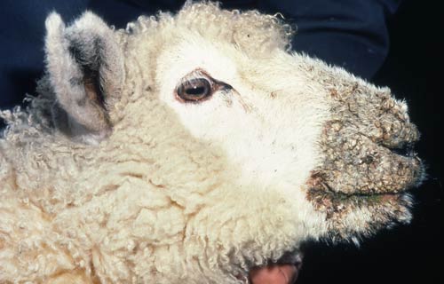
ORF
Clinical symptoms
- Appearance of nodular eruptions on the oral commisures, lips, mouth and nostrils and the lesions are followed
by papules, vesicles, pustules and ulcers in 3 to 4 days.
- Extensive lesions on the feet lead to lameness.
- Mastitis may result in ewes with lesions on the udder.
- Profuse salivation, lacrymation accompanied by nasal discharge.
- Ewes nursing infected lambs may develop lesions on the udder.
- In young lambs, the initial lesion may develop on the gum below the incisor teeth.
Suggested first aid
- Affected animal should be segregated from the rest of the flock.
- Strict hygienic and sanitary measures are to be adopted.
- Contact nearest Veterinary Assistant Surgeon for treatment.
General control and preventive measures
- Lambs should be vaccinated when one month old.
- For better results, a second vaccination 2-3 months later is suggested.
- Vaccines should be used cautiously to avoid contaminating uninfected premises and vaccinated animals should be
segregated from unprotected stock until the scabs have fallen off.
- Strict hygienic and sanitary measures are to be adopted.
- Non immunized lambs should be vaccinated before entering infected feedlots.
ANTHRAX
Symptoms
- Sudden death within 48 hrs of illness of animal
- Following death, there is oozing of blood from the natural orifices.
- Bloat may develop
- Oedema may predominantly notice under the neck, brisket region, thorax, abdomen and flank.
Suggested first aid
- The dead animal body should not be opened.
- Should have consultation with nearest qualified veterinary doctor.
- This disease should be brought under the notice of the regulatory officials in case of an outbreak.
- Care should be taken to destroy the dead body by deep burial with quick lime.
Prevention and control
- Periodical and regular vaccination should be done.
- Strict quarantine measures in anthrax prone areas.
- Preventing the introduction of infected animals into disease free areas.
- Care should be taken to destroy the dead body by deep burial with quick lime.
- Persons handling the anthrax infected animals should adopt adequate sanitary measures.
- The adjacent areas of the dead and infected animals should be thoroughly disinfected by 3% per acetic acid or
10% caustic soda or 10% formaline.
- The fodder from infected pasture should be destroyed and not to be given to the other animals.
JOHNE’s DISEASE
• Johne`s disease is a specific chronic contagious enteritis of cattle, sheep, goat, buffaloes and occasionally
of pigs. The disease is characterized by progressive emaciation and in cattle and buffaloes by chronic diarrhea
and thickening of the intestine.
Transmission
- Under natural conditions the disease spread by ingestion of feed and water contaminated by the faeces of
infected animals. The infection occurs mostly in the early month of life.
- The incubation period extends from 12 months to several years. The animal aged 3 to 6 years mostly suffer from
the disease.
- Affected animals may not show clinical symptoms continue to discharge organisms in faeces. The organisms
persist in pastures for about 1 year. The organisms are susceptible to sunlight, drying and high PH of soil;
continuous contact of urine with faeces reduces the life of bacteria.
- The infected animals which are apparently healthy often show clinical signs after parturition.
Treatment
•The organisms are more resistant to chemotherapeutic agents in vitro than Mycotuberculosis. Because of this the
practical utility of treatment in clinical cases is poor.
Control
- The affected animal should be segregated and their faeces properly disposed off.
- Alive vaccine has been developed. It reduces the incidence of clinical disease.
- It consists of a non-pathogenic strain of Jhone`s bacillus with an adjuvant.
- The calves soon after birth are inoculated with vaccine subcutaneously. The vaccinated animals become reactors
of Jhonin. Vaccination is generally done in heavily infected herds.


