BACTERIAL DISEASES
i. ANTHRAX
B.anthracis causes Anthrax in animals. Bacillus anthracis spores remain viable for many years in soil, water and
animal hides and products. The main routes of entry of endospores are by ingestion, from soil when grazing or in
contaminated food and by infection of wounds. Cattle, sheep and goats are most susceptible to infection.
Symptoms
- In peracute sepeticemia death occurs within 2 hours after animal collapsing with convulsions, sudden death in
animals that appeared normal is common.
- In acute septicemia death occurs within 48 to 96 hours clinical signs include fever, anorexia, ruminal stasis,
hematuria and blood tinged diarrhea.
- Pregnant animals may abort and milk production often abruptly decreases.
- Terminal signs include severe depression, respiratory distress and convulsions.
Prevention and Control
Prevention of anthrax in animals is aided by active immunization. The organism is susceptible to penicillin-G,
tetracyclines, erythromycin and chloramphenicol.
ii. HAEMORRHAGIC SEPTICEMIA
Symptoms
- Fever, a sudden drop in milk yield, signs of abdominal pain, severe diarrhoea and dysentery, respiration
becomes rapid and shortly before death the mucous membranes appear cyanotic.
- In less acute cases there will be odema development in the region of the head, neck and brisket. The nasal
discharge may be blood stained or purulent. Death occurs within 2-4 days.
Control and prevention
- Pasteurella is amenable to Penicillin-G, streptomycin, chloramphenicol, chlortetracycline, sulpha and
tripmethoprim, enrofloxacin and oxytetracycline.
- Vaccination
iii. BLACK QUARTER
Clostridium chauvoei causes black quarter or black leg in Cattle. Gram positive, rod shaped with rounded ends. Worldwide
distribution in soil and pastures.
Symptoms
- The disease usually occurs in young cattle of 6 months to about 2-3 years of age. Crepitating swelling in the
hind or fore quarter, lameness, muscles shows trembling with violent twitching. Death usually occurs within 24
hours.
Control and prevention
- Hyper immune serum (HIS) is used to control explosive outbreaks. Penicillin along with HIS is used to treat
the disease.
- Oxytetracycline & Chlortetracycline can also be employed effectively in early stages
iV. BOVINE TUBERCULOSIS
Mycobacterium bovis causes bovine tuberculosis in many animal species and also cause tuberculosis in human
Clinical signs
- In general form, the affected animals become docile, progressive emaciation, capricious appetite, fluctuating body temperature and rough / sleek hair coat, animal does not put up weight. All these general signs are pronounced following calving.
- In respiratory form, silent or paroxysmal cough especially during early morning and chilled weather. Chronic cough with dyspnoea, squeaking crackles , enlargement of retropharyngeal lymphnode causes dysphagia and noisy breathing due to pharyngeal obstruction.
- In reproductive form, metritis and inflammation of placenta leads to infertility, abortion and failure in conception.
Control and prevention
- Treatment and vaccination are inappropriate in control programmes for cattle. In many countries, tuberculin testing followed by isolation and slaughter of reactors has been implemented as the basis of national eradication schemes.
V.BRUCELLOSIS
Brucella abortus species are obligate intra cellular parasites and cause abortion in last trimester of pegnancy
Symptoms
- The disease in cattle is almost always caused by B.abortus.
- The incubation period is usually from 30 to 60 days.
- After bacteraemia the infection localizes in the placentae, if the animal is not pregnant, the infection
localizes in udder (interstitial mastitis).
- In the bull, orchitis and epididymitis.
- Abortion at 6 months and retained placentae are the cardinal signs.
Prevention and control
- The attenuated live vaccine is used in female calves 4 to 12 months of age.
- The adjuvant bacterin is used as booster vaccine.
Vi.BLACK QUARTER
Black quarter is an acute infection but a non-contagious disease characterized by inflammation of muscles,
severe toxaemia and high mortality in cattle and sheep.
Transmission
- In cattle the disease is confined to young stock between the age of 6 months and 2 years.
- Buffaloes usually suffer a mild disease. The outbreaks occur with a onset of rainy season.
- The cattle acquire infection from ingestion of organism and the ingested bacteria remain as dormant spores in tissues until predisposing factors stimulate the development of negative forms and rapid multiplication and formation of toxins.
Symptom
- The most obvious sign is a crepitate swelling in hind- or forequarters crackles when rubbed due to gas in the muscle.
- The symptoms are fever, lameness and switching of the muscles of the affected region.
- Sometimes animal may die without showing symptoms. Death usually occurs within 24 hours of the symptoms first observed.
- The affected region is hot and painful but soon becomes cold and painless, and there is crepitation due to gas. The skin over the affected area becomes dry, hard and dark.
Control
- Hygiene and prophylaxis are the methods of control.
- Proper hygiene requires the destruction of carcases by burning, and cleaning and treatment of all wounds.
- Active immunization of animals is proved to be effective. The vaccine used is formalized alum precipitated whole culture vaccine. It is a common practice to vaccinate animals before the onset of rainy season.
vii. JOHNE`S DISEASE
Johne`s disease is a specific chronic contagious enteritis of cattle, sheep, goat, buffaloes and occasionally of pigs. The disease is characterized by progressive emaciation and in cattle and buffaloes by chronic diarrhea and thickening of the intestine.
Transmission
- Under natural conditions, the disease is spread by ingestion of feed and water contaminated by the faeces of infected animals. The infection occurs mostly in the early month of life. The incubation period extends from 12 months to several years.
- The animal aged 3 to 6 years mostly suffer from the disease. Affected animals may not show clinical symptoms and continue to discharge organisms in faeces.
- The organisms persist in pastures for about 1 year. The organisms are susceptible to sunlight, drying and high PH of soil; continuous contact of urine with faeces reduces the life of bacteria.
- In cattle, clinical signs appear mainly during 2-6 years of age. The infected animals which are apparently healthy often show clinical signs after parturition.
Control
- The affected animal should be segregated and their faeces properly disposed off.
- Alive vaccines have been developed. It reduces the incidence of clinical disease. It consists of a non-pathogenic strain of Jhone`s bacillus with an adjuvant.
- The calves soon after birth are inoculated with vaccine subcutaneously. The vaccinated animals become reactors of Jhonin.
- Vaccination is generally done in heavily infected herds.
VIRAL DISEASES
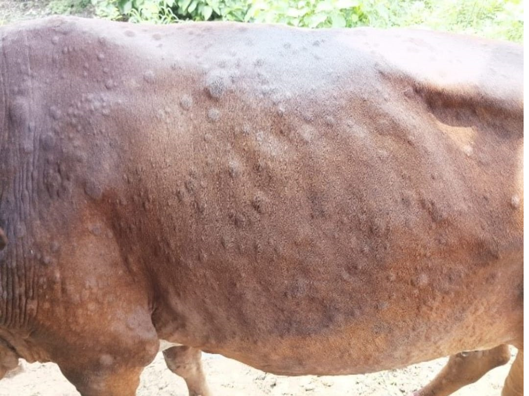
LUMPY SKIN DISEASE IN CATTLE
Lumpy skin disease is an infectious, eruptive, occasionally fatal disease of cattle characterized by nodules on the skin and other parts of the body. Secondary bacterial infection often aggravates the condition.
Etiology
The causal virus is related to that of sheep pox. Lumpy skin disease appears epidemically or sporadically. Its incidence is highest in wet summer weather, but it may occur in winter also. It is most prevalent along water courses and on low ground. The disease spread through insect bites which are suspected as mechanical vectors; however, outbreaks have occurred under conditions in which insects practically could be excluded.
Clinical findings
Infected cattle develop fever, lacrimation, nasal discharge, and hypersalivation, followed by the characteristic eruptions on the skin and other parts of the body in ~50% of susceptible cattle. The incubation period is 4–14 days. The nodules are well circumscribed, round, slightly raised, firm, and painful and involve the entire cutis and the mucosa of the GI, respiratory, and genital tracts. Nodules may develop on the muzzle and within the nasal and buccal mucous membranes.
The skin nodules contain a firm, creamy-gray or yellow mass of tissue. Regional lymph nodes are swollen, and edema develops in the udder, brisket, and legs. Secondary infection sometimes occurs and causes extensive suppuration and sloughing; as a result, the animal may become extremely emaciated, and euthanasia may be warranted. After certain time, the nodules either regress, or necrosis of the skin results in hard, raised areas clearly separated from the surrounding skin. These areas slough to leave ulcers, which heal and scar.
Morbidity is 5%–50%; and mortality is usually low. The greatest loss is due to reduced milk yield, loss of condition, and rejection or reduced value of the hide.
Preventive measures
- Control of animal movement
- Restriction with affected animals and persons dealing with such animals
- Preventive vaccination should also be undertaken in high risk areas like border area of affected district and state and animals should be identified and documented.
Biosecurity measures
- Immediate isolation of sick animal from the healthy animals. Symptomatic treatment of affected animals may be carried out with all precautions and biosecurity measures. Feeding of liquid feed, soft feed and fodder is recommended.
- Clinical surveillance against LSD in affected districts and around surrounding villages should be intensified.
- The buffaloes should be kept separately till complete recovery of the affected animals, if reared together.
- Disinfection of premises at regular intervals.
- Ecto-parasiticide should also be applied to healthy animals on the infected and on surrounding farms.
- The persons dealing with the infected animal should wear gloves and face masks and carry out hygienic and disinfection measures at all times.
- Care should be taken to report any unusual sickness of other animals to the nearest veterinary Hospital/Dispensary.
- Hygiene practices should be followed at the animal farm and by the people in areas where animals are infected.
- Farms with affected animals should be visited regularly by field veterinarians until all the cases are recovered. The veterinary staff should take all precautionary hygiene measures to avoid the further spread of disease to other farms/households.
- In case of mortality, carcass should be disposed of by deep burial method observing all hygienic measures.
- Cattle markets located within 10 km radius of the epicentre of infection should be closed.
- Trade of live cattle, participation in fairs, and shows should be banned immediately upon confirmation of the disease in the affected areas.
- Semen from LSD-affected animals should not be collected and processed for production and distribution.
- Control of vector population (ticks, flies, mosquitoes, fleas, midges) in the premises and the animal body should be carried out using insecticide, repellents and other chemical agents.
- Affected Premises, vehicles plying through the affected animal holdings should be carried out with appropriate chemicals/disinfectants [Ether (20%), chloroform, formalin (1%)].
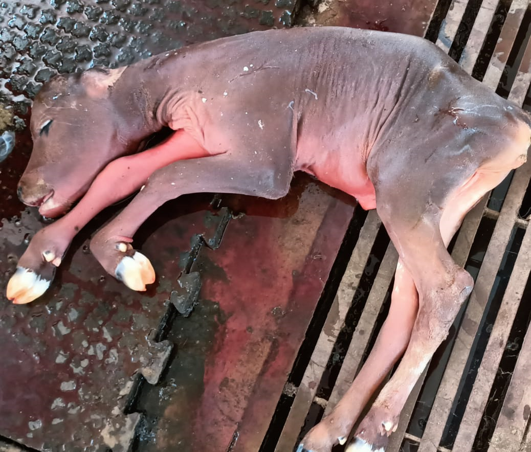
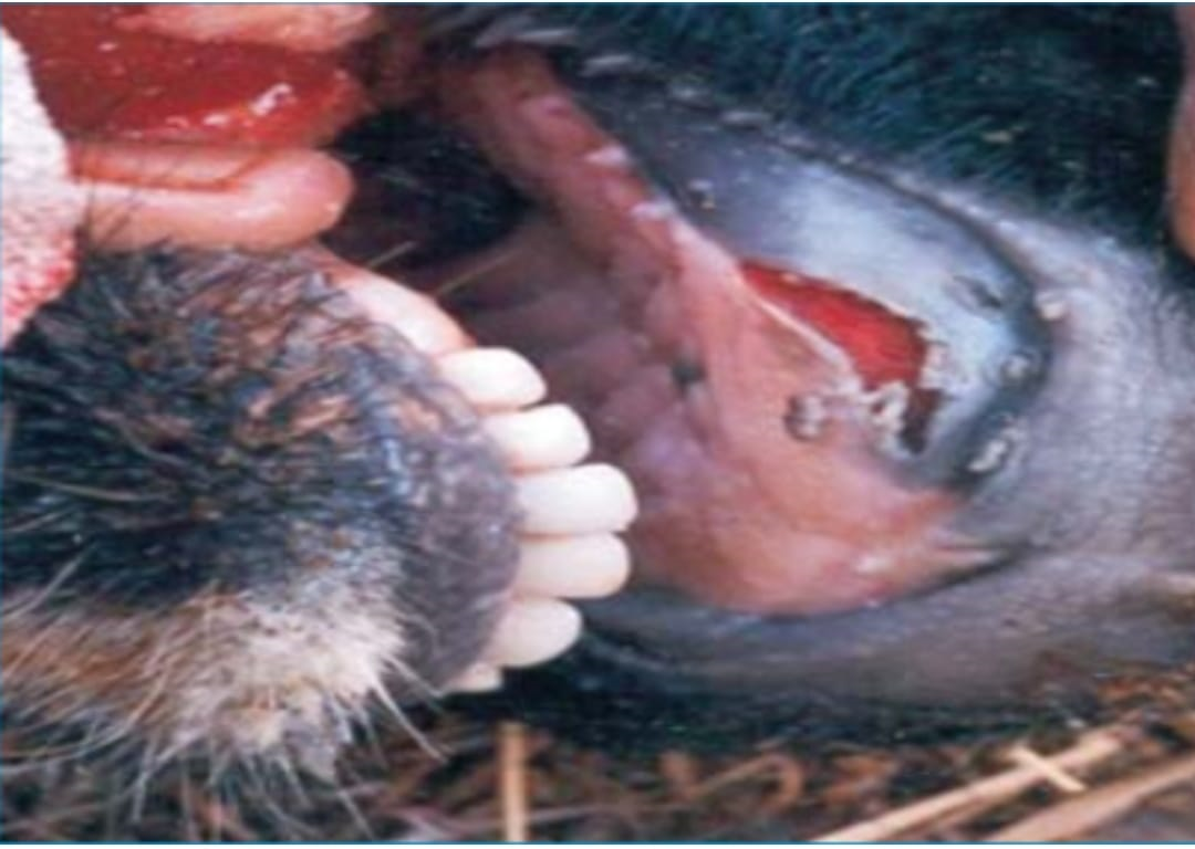
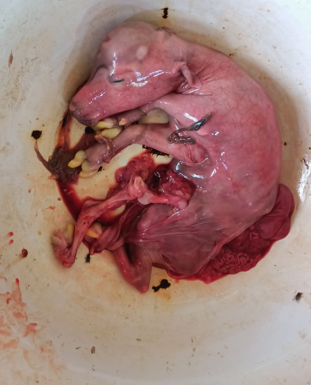
FOOT AND MOUTH DISEASE
Foot and mouth disease (FMD) is the most contagious disease of mammals and cause severe economic loss in susceptible cloven-hoofed animals (cattle, pigs, sheep, goats, and water buffalo).
The transmission is through direct contact, through water, manure, pasture and cattle attendant
Symptoms
The disease is characterized by the formation of vesicles (fluid-filled blisters) and erosions in the mouth, nose, teats and feet. Initial signs are pyrexia (39.4-40.6ºC), dullness, anorexia, and fall in milk production. These signs are followed by excessive salivation; drooling, serous nasal discharge; shaking, kicking of the feet or lameness; and vesicle (blister) formation in the tongue, dental pad, gums, soft palate, nostrils, muzzle, interdigital space, coronary band, and teats. Pregnant cows may abort, and young calves may die without developing any vesicle. The course of an FMD infection is 2 to 3 weeks. Secondary infection may delay recovery.
Prevention & Control :
- Thorough disinfection of shed, utensils, clothes of attendants.
- Vaccination – polyvalent – once – 4months or varies with type of vaccine
BUFFALO-POX
The disease occurs in India in both generalized and localized forms, udder, inner thigh, lips and nostrils. The disease is of zoonotic importance manifesting lesions on the hands and fingers of milkers. The methods of treatment and prevention are similar to those recommended for cow-pox. Since buffaloes wallow in marshy places, care should be taken to see that the wounds are cleaned well and kept free from flies. Attempts to develop a vaccine against buffalo-pox have not given encouraging results.
Symptoms
- After an incubation period of 2 to 5 days, there is some rise in body temperature; the animal develops pin-point red spots and papules of the size of mustard or sago which can be felt by hand.
- Later, these papules coalesce into vesicles, papules occurring on the udder are generally circular, but those on the teats are elongated.
- The lesions heal in the course of 15 to 20 days; the udder and the teats regain their normal appearance.
- In males, the disease is very often unnoticed, as scrotum potion and inside region are often covered with dirt and consequently hidden from view.
Treatment, Prevention and Control
- The lesions heal by themselves in the normal course and the adoption of special measures is not called for; only the usual rules of hygiene need to be observed.
- The lesions should be cleaned with a 1:1,000 solution of potassium permanganate followed by the application of an antiseptic ointment such as 1:110 boric acids.
- The affected animals should be isolated and milked by separate milkers. Milk from affected animals should be boiled before use.
- If the disease assumes serious proportions, vaccination may be undertaken by scarification in the perineum with calf lymph or with material collected from lesions from the animal.
METBOLIC DISEASES
Milk fever
Milk fever is a metabolic disease in cows soon after calving due to fall in serum calcium level in cows after calving as a result of failure to mobilize calcium reserves and of the development of negative calcium balance in late pregnancy.
Symptoms
- Disease flares up within 72 hours of calving. Initially the cows show excitement, in coordination of movement muscular tremors in limbs and head, lying in recumbent position with her head directed towards flank. In final stages, subnormal temperature, dilatation of the pupil, impalpable pulse, coma and death is noticed.
Control
- Continued mixing of ½ liter of supernatant lime water for cow may reduce the incidence.
- Optimum quantity of Calcium and Vitamin D3 are advised
BLOAT: (TYMPANY)
Bloat is a disease of ruminants in which rumen and reticulum is over distended with the gases of fermentation.
Cause and Symptoms
- Excess intake of fresh legumes and feeding of high grain ration lead to frothy bloat. Obstuction to normal expulsion of gases from rumen by choking the esophageal passage by corncob, turnip and sugar beet cause free gas bloat.
- Acute form of tympany results in sudden death before rendering any aid to the affected animal. In acute cases, the distension of the rumen occurs quickly, the flank and the whole abdomen is enlarged. On percussion, the left flank produces a drum like sound, initially the animal frequently gets up and lies down, kicks at belly and even rolls. Breath becomes difficult and is evidenced by oral breathing, protrusion of tongue and salivation.
- When the distension of abdomen becomes extreme, the animal exhibits uncoordinated movement, inability to stand, falls all of a sudden. Collapse and death occur quickly.
- In chronic tympany, the distension of abdomen and intra-abdominal pressure are not serious. The gas is ‘free’ but retained because of obstruction of the passage thereby preventing normal eructation of gases.
Diagnosis
- Based on characteristic symptoms of distension of abdomen and distress by the affected animal.
Control and Treatment
- In per acute cases, puncture the rumen with a sharp knife or with a trocar and canula to expel the gases. Administer orally oil of turpentine 60 ml well mixed with one litre of groundnut oil or ginger oil or cocounut oil. After six to eight hours, administer powdered ginger 30 grams, Asafoetida 30 gram, well mixed to jaggery.
- Fresh legumes should be wilted and then fed to stall fed animals. Feed dry roughages before turning the cattle to luxuriant pasture to avoid bloating.
MASTITIS
Mastitis is an inflammation of the mammary gland in which the milk undergo physical, chemical and microbiological changes where as mammary glandular tissue undergo physical and pathological changes.
Etiology and Clinical signs
- Mastitis is caused majorly by Staphylococcus, Streptococcus and coliform bacteria and less importantly by other organism such as other bacteria, viruses, and fungus.
- Per acute form: Pyrexia, anorexia, respiratory distress, swollen, hot and painful udder. Cessation of milk production. Exudate are often blood stained.
- Acute form: Swollen udder, changes in quality of milk. Milk becomes curd like, yellow, brown fluid with flakes and clots.
- Subacute form: No changes in the udder tissue.
- Chronic form: Udder is haemorrhagic, and fibrotic. Swollen and palpable supra mammary lymphnode,. Udder is thick, firm, nodular and atrophic, yellowish or white fluid with clots and flakes.
Treatment
- Stripping out the milk from the infected quarters. Cleaning of infected quarters with normal saline and distilled water. Infusion of antibiotic therapies immediately after the infection. Continuous use antibiotics as per the antibiogram.
Control:
- Hygenic measures are important.
- Animals diagnosed positive should be milked at last.
- Milkers should wash their hands before milking and should use well washed utensils.
- A separate clean cloth for each cow is used for washing the udder with a disinfectant.
- The first stream of milk from each quarter should not be allowed to drop on floor but collected in a separate container.
- Milkers should not wet their hands with first stream of milk.
PARASITIC DISEASES
Anaplasmosis
- Anaplasmosis is a vector-borne, infectious blood disease in cattle caused by the rickesttsial parasites
Anaplasma marginale and Anaplasma centrale.
- It can also be transmitted via contaminated needles, dehorning equipment, castrating knives, tattoo
instruments, biting flies and mosquitoes.
- The intracellular parasite destroys red blood cells. It causes anemia, fever, weight loss, breathlessness,
uncoordinated movements, abortion and death.
Bovine Babesiosis(Red water disease, Tick fever)
- Bovine babesiosis is a febrile, tick-borne disease of cattle and buffalo, caused by one or more protozoan parasites of the genus Babesia.
- The acute form is generally characterized by rapid growth and multiplication of the parasite in blood with extensive erythrocytic lysis leading to anemia, icterus, hemoglobinuria, enlargement of the spleen, and frequently, death.
- The term "Babesiasis" refers to the subclinical and chronic infections that usually persist following recovery
from initial attack by the parasite.
- The chronic form is poorly defined clinically and is associated with anemia and variable weight loss.
Theileriosis
- Theileriosis is a disease of mammals- caused by T. parva and T. annulata in cattle
- Marked pyrexia lymph node enlargement, dyspnoea, epistaxis, emaciation, diarrhoea, and other GI signs.
- Ocular signs and masses may develop.
- Pruritus and skin lesions/plaques are also seen.
- Neurological and reproductive signs may develop in chronic or endemic disease.
- The degree of pyrexia, pathogen load and host susceptibility will determine the severity of clinical signs at presentation.
REPRODUCTIVE DISORDERS
1.Repeat breeding
A cow with normal oestrous cycle which has failed to conceive within 3 or 4 consecutive services is called as Repeat breeders.
Causes
- Genetic /Anatomical defects in genital tract
- Sperm abnormalities viz. knobbed sperm, Dag sperm
- Abnormalities in ovum viz. degenerative changes, rupture or shrinkage of ovum.
- Ageing of sperm or ova
- Infection of the female genital tract (Endometritis) results into early embryonic death.
- Nutritional factors - Deficiency of vitamins and minerals.
-
Managerial Factors
- Stress of Long Distance Transportation, High Temperature
- Failure to Detect Heat in Proper Time
- Improper Storage of Semen
- Improper Thawing of Frozen Semen
- Improper Insemination Technique
- Improper Timings of Inseminations
- Details about each managerial factor.
Precautions to check repeat breeding problem
Repeat breeding is very widespread problem of dairy cattle which causes economic loss to the farmers. As prevention is better than cure, the following precautions may be taken to minimize the incidences of repeat breeding in dairy animals.
- Maintain breeding record properly
- Avoid overcrowding especially at the time of AI.
- Mineral mixture supplementation should be an integral part of diet @ 2% of ration.
- Provide clean water to drink.
- Provide as much as cool climate to the animals during summer especially to crossbred animals. Heavy plantation around the farm, sprinkler water, bathing and roof painting will help to keep the animals cool and healthy in summer.
- Underweight (less than 250 kg), malnourished, anaemic animals should never be inseminated.
- Overfeeding of oil seed cakes to dairy cows should be avoided since it can enhance embryonic mortality.
- Do not feed mould infested grains and green fodder and soiled wheat straw to dairy cows.
- Cows showing turbid or discoloured mucus discharge should not be inseminated and should be checked for any uterine infections and treated accordingly.
- Avoid putting the straw back into the liquid nitrogen cylinder once after it has been taken out. Once straw has been taken out, it should be used immediately or thrown away.
- To avoid contamination, AI gun should never be pulled out of the vagina and reinserted into it.
- Only efficient trained personnel should do artificial insemination.
- Never use bulls with known genital infections for AI.
- Do not use same bull again and again for breeding in same cow.
- Use hormonal treatments judiciously only when required.
2.Prolapse of Vagina and Uterus
It is defined as an eversion of genital organs. It is seen either before or after parturition.
Etiology
- Inherited tendency
- Low levels of progesterone
- Urogenital infections like cervicitis and vaginitis
- Dystokia
- Breeding injury
- Retention of placenta
- Straining due to diarrhoea or constipation and consumption of estrogen rich plants or feeds
Clinical signs
- Protrusion of uterus, cervix and/or vagina beyond vulva
- Continuous straining
- Wounds or injury on the prolapsed mass
- Restlessness / uneasiness
- Rise in body temperature
- Loss of appetite and death in severe cases.
Care and Prophylaxis
- Washing of prolapsed mass with antiseptic solution.
- Application of antiseptic ointment on prolapsed part.
- Reduction of protruded part by cold fomentation.
- Reposition of the prolapsed part manually.
- Apply rope-truss to provide support and for retention.
- Keep the animal in slanting position with hindlegs at higher level and head at lower level.
- Eliminate causes of irritation or straining
- Avoid injury or unnecessary traction at delivery
- Early treatment of retention of placenta cases